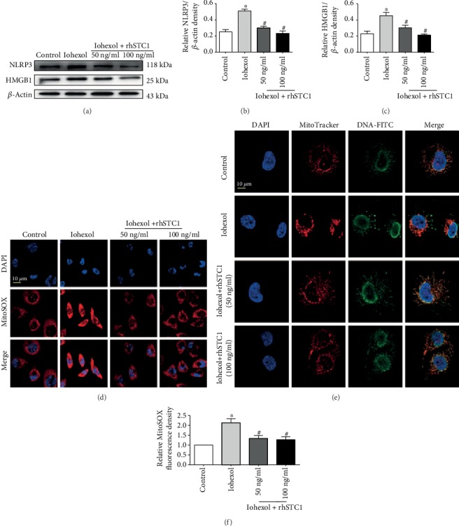
Figure 2.

rhSTC1 treatment attenuates inflammation injury and mitochondrial ROS generation in HK-2 cells. HK-2 cells were treated with iohexol (200 mg I/ml) with or without rhSTC1 at different concentrations (50 ng/ml, 100 ng/ml) at indicated time courses, respectively. (a) Western blot analysis of the expression of HMGB1 and NLRP3 (n = 3). (b, c) Quantification of average Western blot band intensities. (d–f) Representative images of mitochondrial ROS generation (d) and mitochondrial DNA release (e) (n = 3). DAPI staining was performed to label nuclear (blue). Mitochondrial ROS was labeled by MitoSOX (red). Mitochondrial DNA was labeled by the indicated antibody (green). Fluorescence images were taken by a LSM780 confocal microscope (magnification, ×630). Values were presented as mean ± SE. ∗p < 0.05, compared with the control group. #p < 0.05, compared with the iohexol group.
