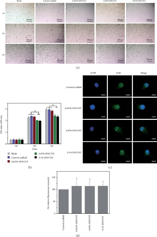
Figure 7.

Proliferation of RSC96 cells. (a) Observation of RSC96 cell proliferation in different culture conditions under a phase contrast microscope. (b) RSC96 cell proliferation measured by CCK8 assay, and the result showed that the scaffolds with 0.05% MWCNT and 0.1% MWCNT demonstrated cytotoxicity on day 3 and day 5 (∗P < 0.05). (c) showed the neurofilament images of RSC96 cells cocultured with scaffolds by immunofluorescence. Scale bar = 50 μm. (d) showed the relative expressions of NF through cell immunofluorescence intensity on scaffolds with different concentration of MWCNTs and compared with the control scaffolds after cultured for 3 days. No difference was found between any two scaffolds.
