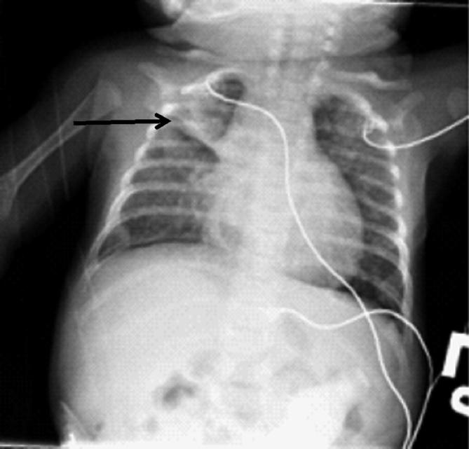
Fig. 7.6.

This chest radiograph, from a young infant with RSV bronchiolitis, demonstrates the typical radiographic features of bronchiolitis including hyperinflation, flattening of the diaphragms, and areas of atelectasis, visible here as a linear density in the right upper lung (arrow)
