Figure 6.
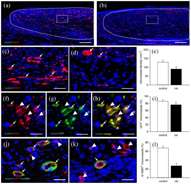
Final vascularization of MVF-seeded matrices. (a–d) Immunohistochemical detection of CD31+ microvessels within MVF-seeded matrices in a PBS- (a, c) and a clo-treated (b, d) mouse (a, b = overview of the implants; c, d = higher magnification of inserts in a and b; broken lines = implant borders; arrows = CD31+ microvessels). Scale bars: a, b = 230 µm; c, d = 45 µm. (e) Microvessel density (mm−2) within MVF-seeded matrices in PBS- (control; white bar; n = 8) and clo-treated (black bar; n = 8) mice. Means ± SEM. *p < 0.05 vs control. (f–h) Representative immunohistochemical detection of CD31+/GFP+ microvessels (arrows) and CD31+/GFP− microvessels (arrowheads) within a MVF-seeded matrix of a clo-treated mouse. Scale bars: 30 µm. (i) GFP+ microvessels (%) within MVF-seeded matrices in PBS- (control; white bar; n = 8) and clo-treated (black bar; n = 8) mice. Means ± SEM. (j, k) Immunohistochemical detection of CD31+ microvessels with (arrows) and without (arrowheads) a perivascular α-SMA+ cell layer within MVF-seeded matrices of a PBS- (j) and a clo-treated (k) mouse. Scale bars: 18 µm. (l) α-SMA+ microvessels (% of all blood vessels) within MVF-seeded matrices in PBS- (control; white bar; n = 8) and clo-treated (black bar; n = 8) mice. Means ± SEM. *p < 0.05 vs control.

