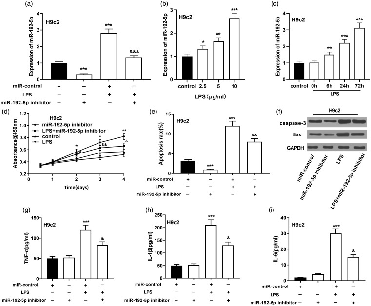
Figure 4.
Effect of miR-192-5p inhibition on the viability and apoptosis of H9c2 cardiomyocytes. (a) qRT-PCR was utilized to measure miR-192-5p in LPS-induced H9c2 cardiomyocytes. (b) qRT-PCR was adopted to work out miR-192-5p in H9c2 cells at different LPS concentrations. (c) MiR-192-5p in H9c2 cells were detected by qRT-PCR at different treatment times. (d) CCK-8 assay was conducted to delve into the viability of LPS-induced H9c2 cardiomyocytes after inhibiting miR-192-5p. (e) Flow cytometry analysis was operated to detect the apoptosis of H9c2 cardiomyocytes induced by LPS after miR-192-5p inhibition. (f) Western blot was conducted to detect caspase-3 and Bax after inhibition of miR-192-5p in H9c2 cardiomyocytes. (g–i) ELISA was carried out to quantify TNF-α, IL-1β and IL-6 expressions in the cell supernatant. vs. miR-control: *, **, *** indicated P < 0.05, P < 0.01, P < 0.001, respectively. vs. LPS group: &, &&, &&& indicated P < 0.05, P < 0.01, P < 0.001, respectively.

