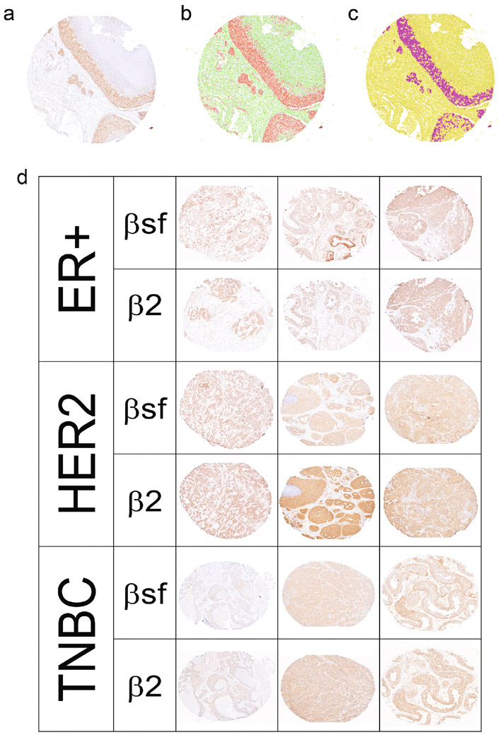
Fig. 6. ERRβ isoform expression by IHC.
A-D. Automatic scanning and semi-automatic quantification of ERRβ splice variant specific mouse monoclonal antibodies (ab) on tissue microarray (TMA). a. Blue/brown staining with ERRβ ab. Automatic detection of b. nuclear v cytoplasmic staining, and c. cells “positive”/ above threshold vs. “negative”/ below threshold. d. ERRβ protein expression in breast tumors. Representative images of ER+, HER2, and TNBC tumor tissues from 150 patient tissue microarray series stained with ERRβ ab

