
Fig. 3.
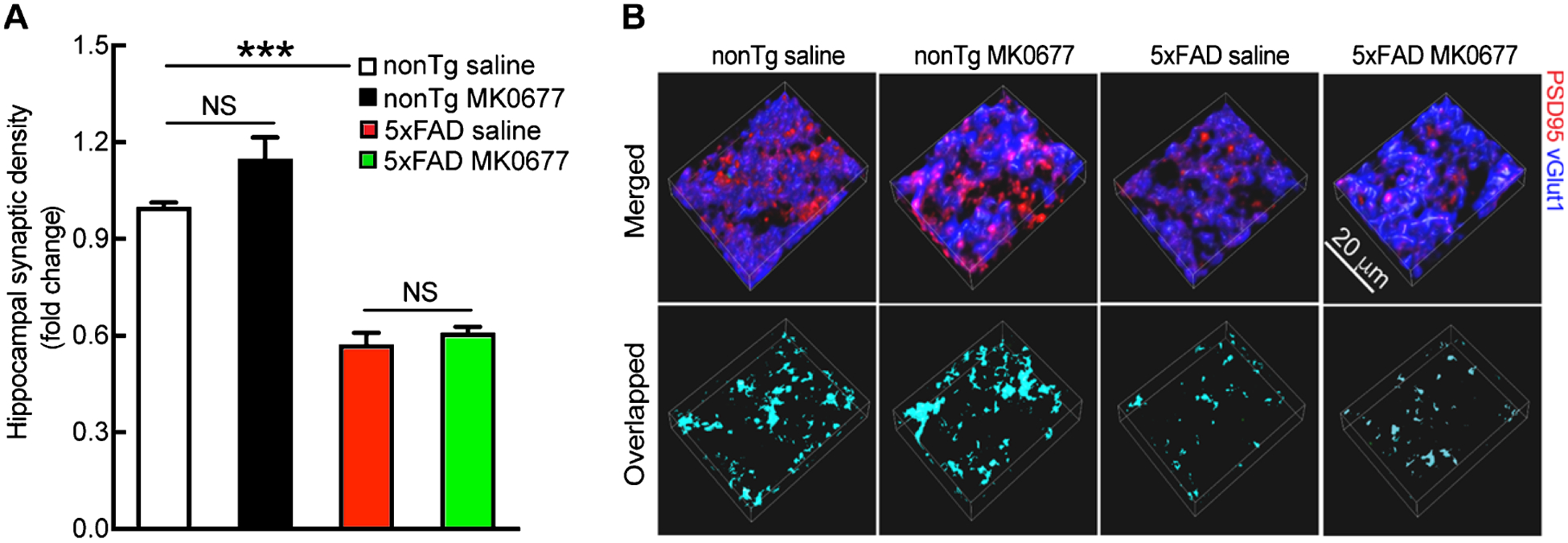
Synaptic density remained unaltered in 5xFAD regardless of MK0677 treatment. A) Saline and MK0677-treated 5xFAD mice showed similar synaptic density. Two-way ANOVA followed by Bonferroni post hoc analysis. t = 7.725, ***p < 0.001 between nonTg saline and 5xFAD saline groups; t = 2.704, NS, no significant difference for nonTg saline and MK0677 treatment groups; t = 0.6671, NS, no significant difference for 5xFAD saline and MK0677 treatment groups. n = 4 mice per group. B) Representative images of synapse staining. vGlut1 (blue) and PSD95 (red) were used as pre- and post-synaptic markers, respectively. The overlaid staining of vGlut1 and PSD95 indicates synapses. Scale bar = 20 μm.