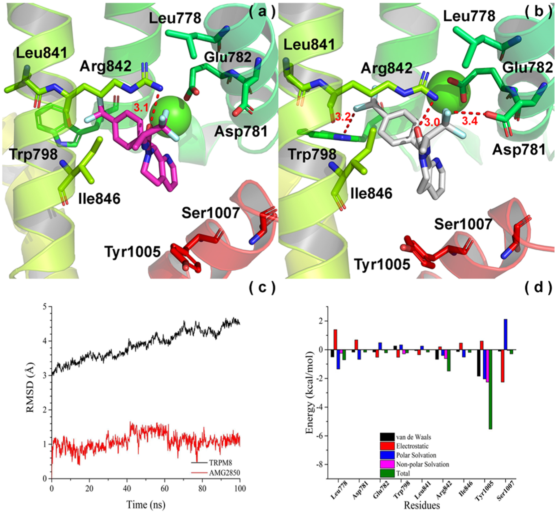
Figure 3.

Convergence parameters of AMG2850 and hTRPM8 during MD simulation. (a) Docking pose (pre-MD) of AMG2850 in hTRPM8. AMG2850 is shown as magenta sticks. Important residues are shown as sticks. Individual helices are colored as follows: S2 (faded green), S3 (green), S4 (yellow-green), TRP helix (red). The Ca2+ ion (green) is shown in CPK representation. (b) Binding mode (post-MD) of AMG2850 in hTRPM8 after 100 ns MD. AMG2850 is shown as gray sticks. (c) RMSD of AMG2850 and hTRPM8 during MD simulation. (d) Energy decomposition of key residues in hTRPM8 that contributed to the binding of AMG2850. Our hTRPM8 homology model was constructed using the cryo-EM structure of TRPM8FA (PDB 6BPQ) as a template.
