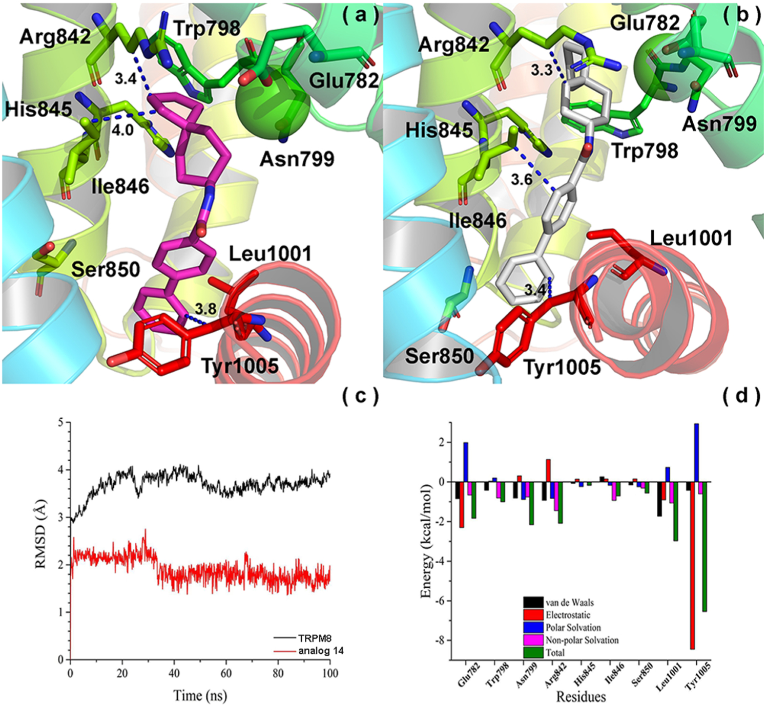
Figure 8.

Convergence parameters of 14 and hTRPM8 during MD simulation. (a) Binding mode (pre-MD) of 14 in hTRPM8. 14 is shown as magenta sticks. Important residues are shown as sticks. Individual helices are colored as follows: S1 (blue), S2 (faded green), S3 (green), S4 (yellow-green), TRP helix (red). The Ca2+ ion (green) is shown in CPK representation. (b) Binding mode (post-MD) of 14 in hTRPM8. (c) RMSD of 14 and hTRPM8. (d) Energy decomposition of key residues in hTRPM8 that contributed to the binding of 14. Our hTRPM8 homology model was constructed using the cryo-EM structure of TRPM8FA (PDB 6BPQ) as a template.
