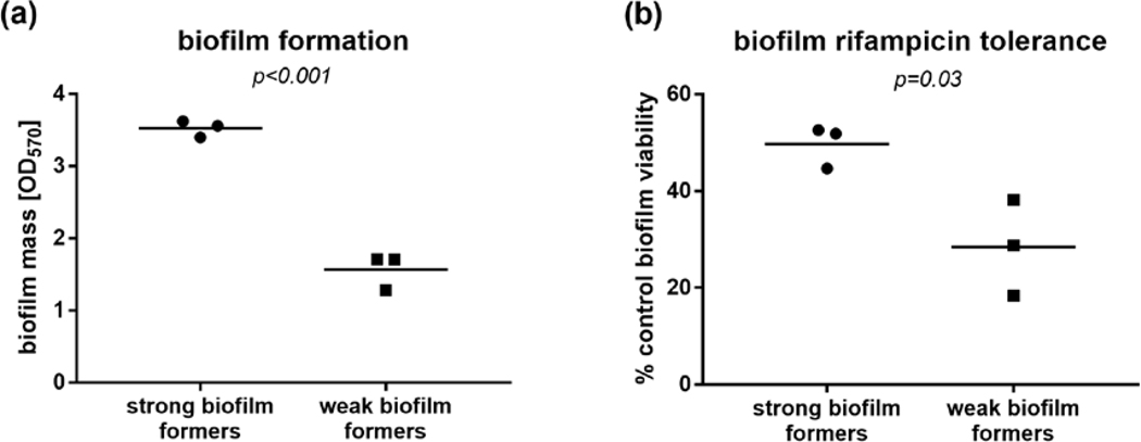
Figure 1.
Biofilm formation in the in 96-well plate crystal violet assay (a) and viability after 24 h exposure to rifampicin, measured with XTT assay (b) of representative ‘strong’ and ‘weak’ biofilm-forming S. aureus strains from the invasive isolate collection. P values calculated with the unpaired t test.

