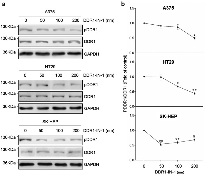
Figure 3.

DDR1-IN-1 blocks of DDR1 phosphorylation in A375, HT29 and SK-HEP cells. Cells were cultured in serum-free basal media. Some cells received exogenous collagen type I and/or the chemical inhibitor of DDR1 phosphorylation DDR1-IN-1. (a) Representative Western blot analysis of phosphorylated (pDDR1) and total DDR1 expression in cells incubated with collagen I and treated with increasing concentrations of DDR1-IN-1. (b) Computer-assisted semi-quantification of the Western blot for pDDR1 and total DDR1 expression in cells incubated with collagen I and DDR1-IN-1, expressed as a ratio pDDR1/total DDR1. Dotted lines indicate the 50% inhibition of DDR1 phosphorylation (IC50). Data are presented as the means ± standard error, n = 2 (*P < 0.05, **P < 0.001).
