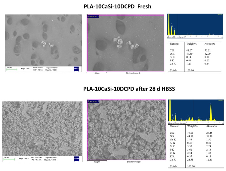
Figure 2.
Surface micromorphology of PLA-10CaSi-10DCPD before and after 28 days immersion in simulated body fluid (500×–1000× magnification); pores of different size (diameter range between 20 and 100 µm) were observed: CaSi and dicalcium phosphate dihydrate (DCPD) microgranules are diffused onto all the structure. In some areas, agglomeration of these granules were also observed. Samples soaked in HBSS for 28 days revealed a well-evident electron dense mineral layer on the materials with pores of approx. 100 µm diameter filled by this layer. Energy dispersive X-ray (EDX) microanalysis on samples not immersed in HBSS showed the polylactic acid (PLA) structural peaks (C and O) and low traces of mineral filler constitutional peaks (Ca, Si, and P); EDX revealed after 28-day immersion in simulated body fluid a consistent decrease of C (from PLA matrix), and a significant elevation of Ca, Si, and P.

