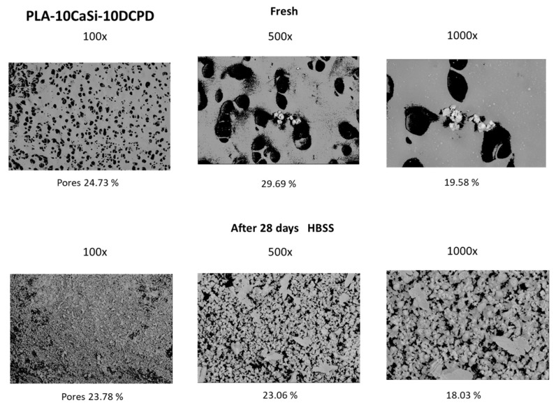
Figure 3.
Mineral-doped surface micromorphology by environmental scanning electron microscope (ESEM) of PLA-10CaSi-10DPCD scaffold. Differences in surface porosity were observed when considering sample immersed for 28 days in simulated body fluids, attributable to calcium phosphate mineral layer deposition which partially covers the polymeric porous matrix.

