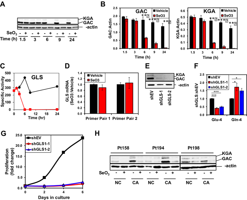
Figure 3. Selenite attenuates GLS expression in A549 cells and human NSCLC tissues.
A) Time-dependent suppression of GLS1 proteins by selenite in A549 cells. A549 cells were treated with or without 6.25 μM Na2SeO3 and harvested at 0, 1.5, 3, 6, 11, and 24 h after treatment for Western blot analysis. Shown is a representative blot. B) Quantification of GAC and KGA intensities in Western blots normalized to that of β-actin p<0.001 *** p<0.01 ** . C) Time course inhibition of GLS activity by selenite in A549 cells. D) Lack of selenite effect on GLS1 gene expression. A549 cells were treated with or without 6.25 μM Na2SeO3 and harvested at 24 h after treatment. Total RNA was extracted and GLS1 gene expression measured using qRT-PCR. ΔΔCt values were calculated with 18S rRNA as housekeeping gene and the ratios of these values for selenite versus control samples were shown for two primer sets. E) Silencing of GLS1 protein expression in A549 cells using two separate shRNA vectors (shGLS’s). Western blot analysis of lysates showed knockdown efficiency for GLS1 shRNA’s against the empty vector (shEV). F) Inhibition of 13C-4-Gln converstion to 13C-4-Glu in 13C5,15N2-Gln-treated GLS1 knockdown cells for 24 h. 13C labeled Gln and Glu were quantified using 1H{13C} HSQC NMR as in Fig. 2. *q<0.05; ***q<0.001; n=3. G) Growth inhibition by GLS1 suppression in A549 cells. H) Suppression of GLS1 proteins by selenite in CA and adjacent NC lung tissue slices. Three matched pairs of CA and NC were treated with or without 6.25 μM Na2SeO3 for 24 h, extracted for proteins, and analyzed by Western blotting, as described in methods.

