
Fig. 2. Loss of Idol expression inhibits plaque formation in a mouse model of Aβ amyloidosis.
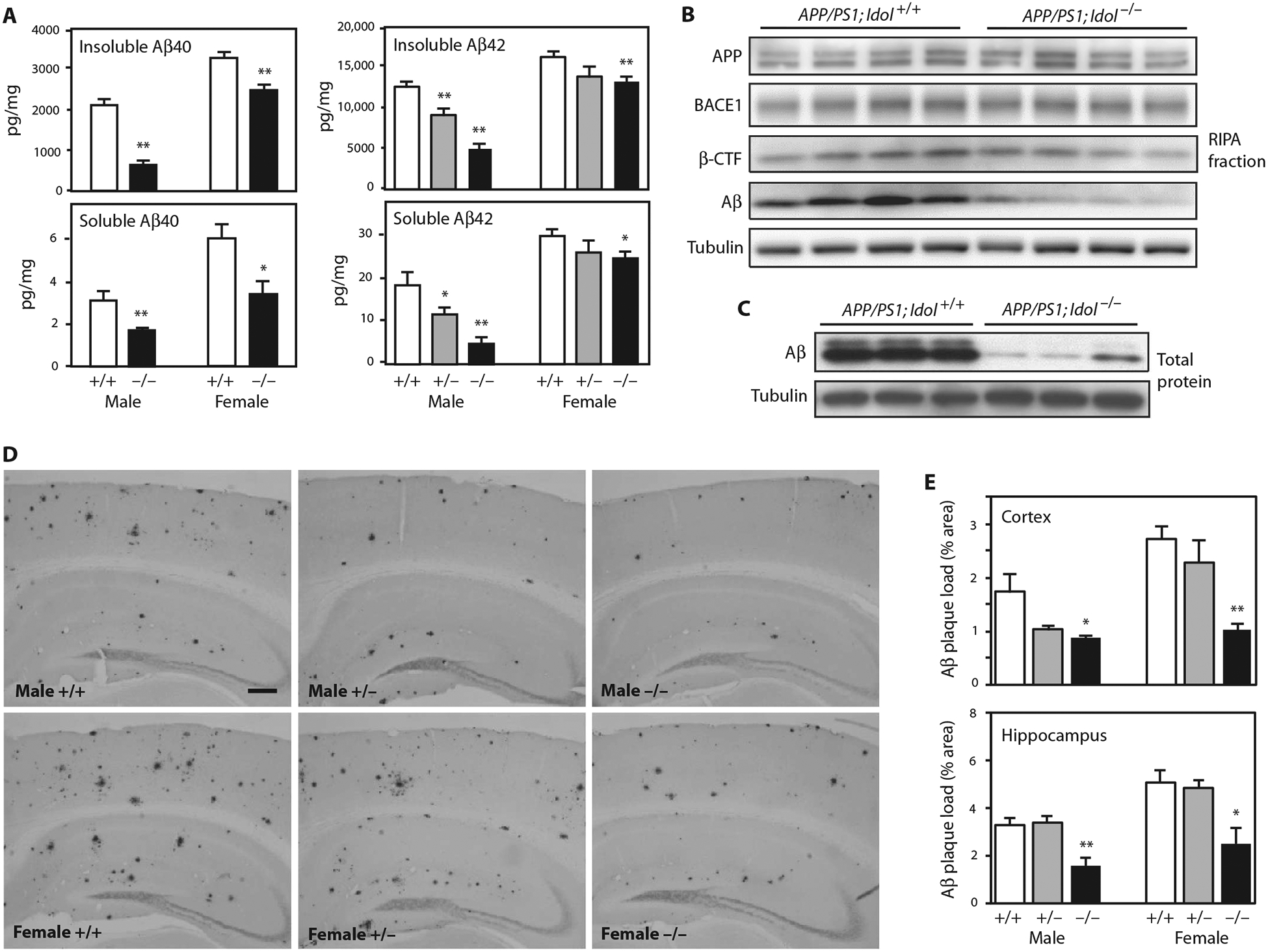
(A) Quantification of Aβ40 and Aβ42 peptides in the insoluble guanidine fraction and the soluble RIPA fraction from the frontal cortex of APP/PS1;Idol+/+, APP/PS1;Idol+/− (for Aβ42), and APP/PS1;Idol−/− mice. Error bars represent SEM. *P < 0.05, **P < 0.01 by Student’s t test. n = 5 to 7. (B) Immunoblot analysis of brain RIPA fractions from APP/PS1; Idol+/+ and APP/PS1;Idol−/− mice. APP was detected with 4G8 antibody. Aβ and β-CTF were detected with 82E1 antibody. (C) Immunoblot analysis of total brain lysates from APP/PS1;Idol+/+ and APP/PS1;Idol−/− mice. (D) Representative micrographs showing immunohistochemical staining of brain sections from APP/PS1;Idol+/+, APP/PS1;Idol+/−, and APP/PS1;Idol−/− mice with Aβ-specific 82E1B antibody. Black stain indicates Aβ plaques. Scale bar, 250 μm. (E) Quantification of Aβ plaque load in the frontal cortex and hippocampus of APP/PS1;Idol+/+, APP/PS1;Idol+/−, and APP/PS1;Idol−/− mice by 82E1B antibody staining for Aβ. Error bars represent SEM. *P < 0.05, **P < 0.01 by Student’s t test. n = 6.