
Figure 2. ZBP1 Senses IAV DVG RNA in the Nucleus.
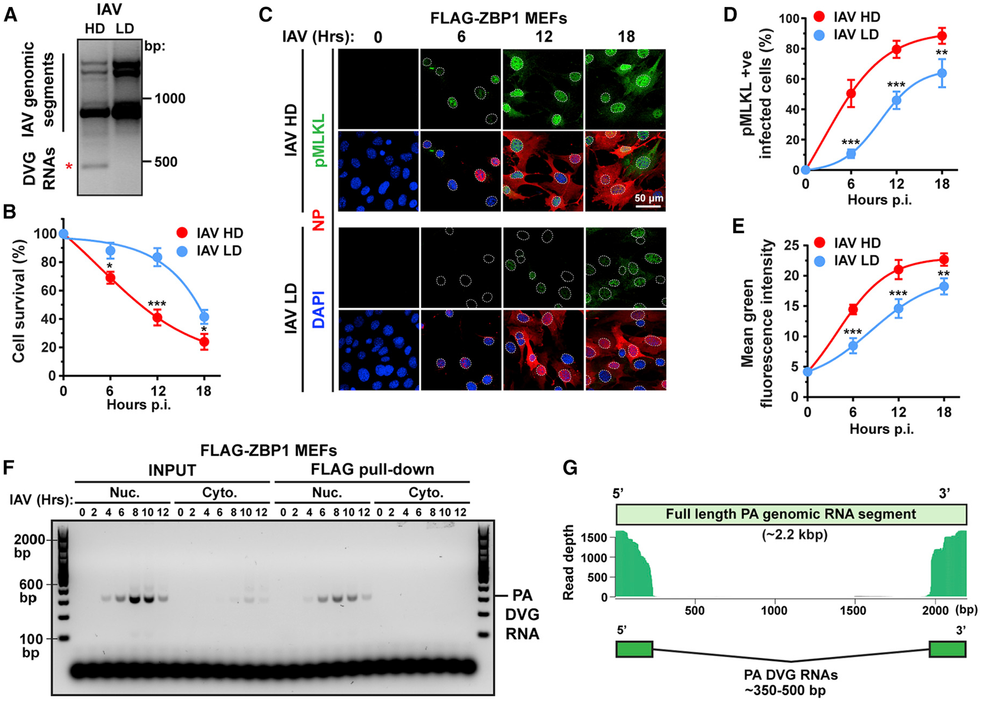
(A) PCR of full-length IAV genomic segments and DVGs in A549 cells infected with IAV PR8 stocks with high (HD) or low (LD) DI particle content. DVGs are indicated by a red asterisk.
(B) Cell death kinetics after IAV HD or LD virus infection of WT MEFs.
(C) Immunofluorescence staining for pMLKL (green) and NP (red) in WT MEFs infected with IAV HD or IAV LD virus. Nuclei are stained with DAPI (blue) and outlined with dashed white lines.
(D) Quantification of pMLKL+ cells as a percentage of infected (NP+) cells.
(E) Quantification of mean fluorescence intensity of the pMLKL signal per cell.
(F) Immortalized Zbp1−/− MEFs stably expressing FLAG-ZBP1 and infected with IAV were separated into nuclear and cytoplasmic fractions at the indicated times p.i. PA segment-derived DVG RNAs in input lysates or eluted from FLAG-ZBP1 pull-downs were determined by PCR.
(G) Paired-end sequencing was performed on PA segment-specific PCR amplicons of IAV RNAs eluted from FLAG-ZBP1 nuclear immunoprecipitates. Sequences with significant overlap are shown mapped to the full-length IAV PR8 PA segment. IAV was used at MOI = 2 in this figure. Data in (A)–(F) are representative of at least three independent experiments. RNA-seq for data in (G) was performed once. Error bars represent mean ± SD. Unpaired Student’s t test, *p < 0.05, **p < 0.005, ***p < 0.0005. Scale bar represents 50 μm.