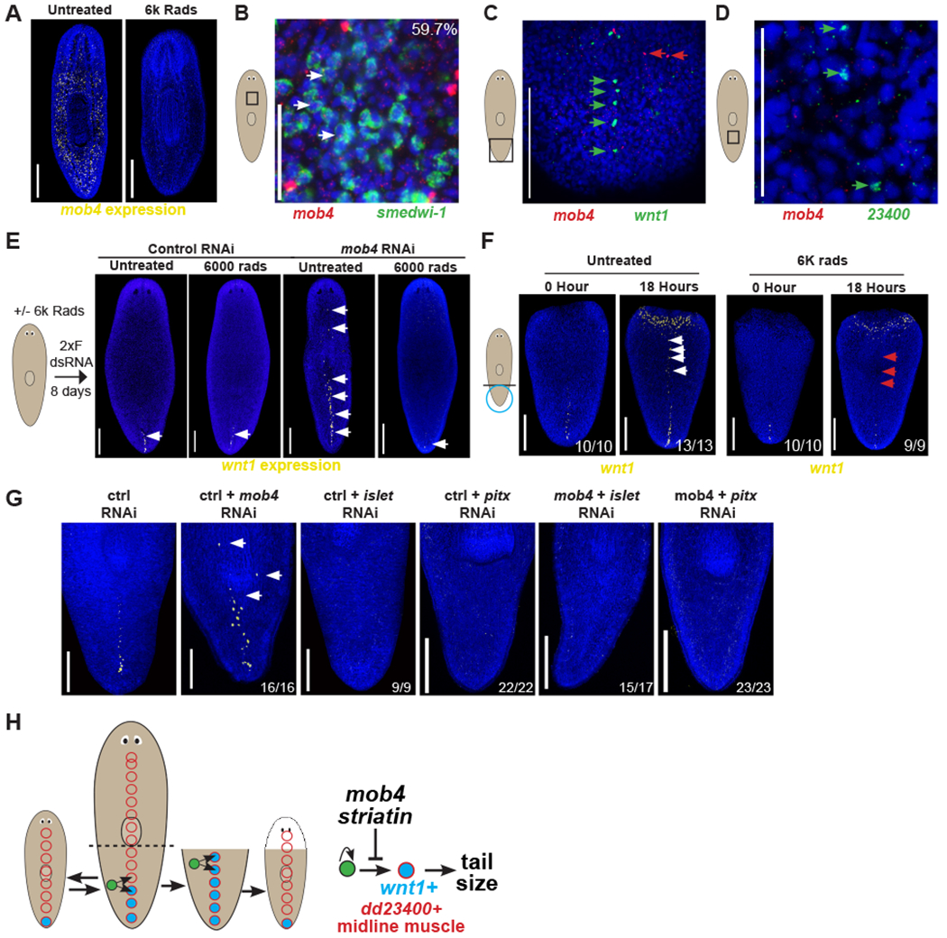
Figure 5. mob4 limits differentiation of stem cells into wnt1+ midline cells.

(A) FISH to detect mob4 showing reduced expression in irradiated animals depleted for stem cells (right) compared to controls (left).
(B) Double FISH of mob4 (red) and smedwi-1 (green) showing mob4 is expressed in 59.7% (242/405) smedwi-1+ stem cells.
(C) Double FISH of mob4 (red) and wnt1 (green) showing mob4 is not expressed in wnt1+ posterior pole cells.
(D) Double FISH of mob4 (red) and dd_23400 (green) showing mob4 is not expressed in dd_23400+ midline muscle cells.
(E) Left 2 images- FISH showing wnt1 expression in untreated vs. neoblast depleted irradiated animals after control RNAi feeding. Right 2 images- FISH showing wnt1 expression in untreated vs. neoblast depleted irradiated animals after mob4 inhibition.
(F) FISH showing expanded wnt1 expression along the dorsal midline in tail fragments at 0 hours or 18 hours after injury in untreated animals (left panels) or treated with 6K rads of X-ray irradiation 2 days prior to surgery. N>5 animals for each time point.
(G) FISH to detect wnt1 showing expanded expression in mob4(RNAi) animals but not in islet(RNAi) or pitx(RNAi) animals. Double RNAi of mob4 with either islet or pitx resulted in the absence of wnt expansion. Bars are 300 microns.
(H) Model showing stem cells (green) giving rise to either wnt1-/dd23400+ (red circle) or wnt1+/dd23400+ pole cells (blue with red border) in the far posterior. mob4/striatin inhibits the differentiation of wnt1+/dd23400+ pole cells. Bars in panels A, E, F are 300 microns and in B-D are 150 microns.
See also Figure S5.
