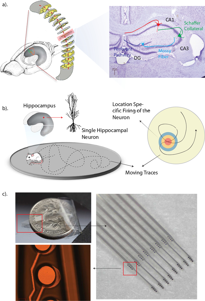
Figure 1.
a). Schematic diagram of the rat brain, the curved hippocampus and a coronal slice of the hippocampus. b). Diagrammatic drawing of the position-specified firing property of hippocampal neurons. c). Photographs of a fabricated, fully functional Parylene multi-electrode array. The photo on top left shows the size of the Parylene array. The right photo shows a zoomed-in view of eight Parylene shanks and sixteen recording groups positioned on those shanks. The bottom left photo shows a representative recording electrode.

