
Figure 2.
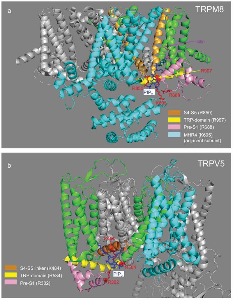
Structure of TRPM8 and TRPV5 with PI(4,5)P2. (a). Structure of TRPM8 with PI(4,5)P2 and icilin (6nr3) from Yin et al [25]. PI(4,5)P2 is colored blue, interacting residues are colored red. Parts of the channel where the interacting residues located are also color-coded: S4-S5 orange, TRP-domain yellow, Pres-S1 segment purple. The remaining parts (S6 and S1-S3) of the same subunit where these residues are located are colored green. The entire adjacent subunit is labeled cyan, the MHR4 region of this subunit contains an additional PI(4,5)P2 interacting residue; most other cytoplasmic parts have been removed for clarity. (b). Structure of TRPV5 with PI(4,5)P2 (6dmu) from Hughes et al [27]. Color-coding is similar to that in panel A.