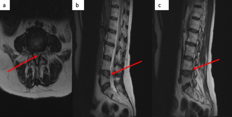
Figure 2. MRI of the spine.
T2-weighted, non-contrast MRI lumbar spine revealing central stenosis at the L4/5 level in the sagittal (b) and axial (a) views with significant thecal sac compression with severe stenosis of the traversing L5 nerve root. The lateral, sagittal view identifies moderate L4/5 foraminal stenosis with impingement on the L4 exiting nerve root (c).

