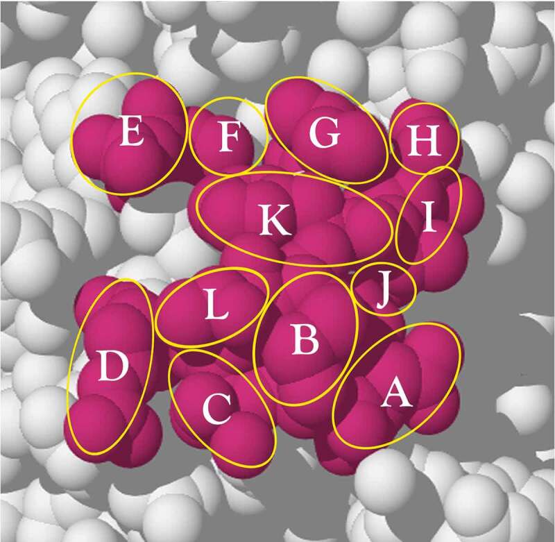
Figure 4.

A surface epitope region (red) consisting of 12 different amino acids selected from the exposed surface of the protein, PCSK9 (PDB: 2PMW). An outline of each residue side-chain is indicated by a yellow circle or oval. The epitope region is used to calculate the combinations of each unique residue set from 1 to 12 residues that could be addressed by different antibodies. The surface region is redrawn from the PDB structure (2PMW; reference 75) using Adobe Illustrator See text for explanation.
