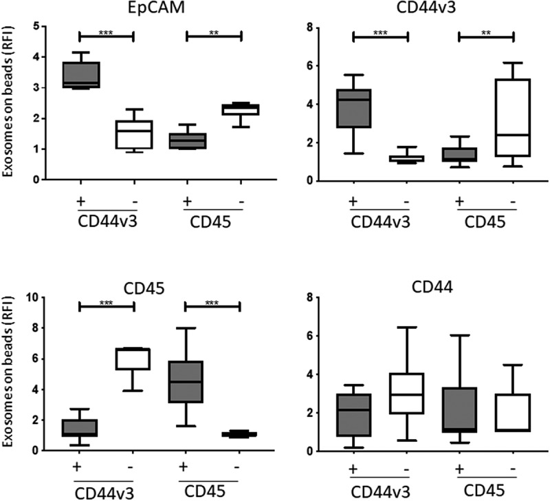
Figure 4.

Detection of antigens carried by immunocaptured CD44v3 exosomes from plasma of HNSCC patients (n = 12). In parallel, exosomes were also captured with anti-CD45 mAb as a control. Flow cytometry-based analysis showed the highest levels of EPCAM on CD44v3(+) exosomes. Expression levels of CD44v3 and EPCAM were also high in the CD45(-) fraction enriched in TEX; they were low in CD45(+) and CD44v3(-) exosomes. CD44 expression levels were comparable in all exosome fractions (*p < .05, **p < .005). Gray bars indicate CD44v3+ or CD45+ exosomes, while white bars indicate CD44V3(-) and CD45(-) exosomes.
