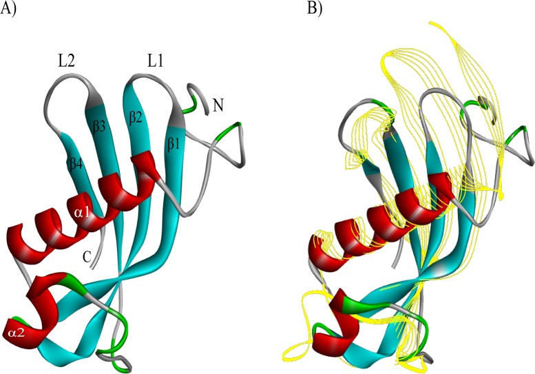
Fig 2. 3D structural comparison of conserved hypothetical protein (Ts_04814) with human cystatin E/M using the I-TASSER program.
(A) The predicted tertiary structure of the conserved hypothetical protein (Ts_04814); there are 2 α-helixes (in red), 4 β-strands (in blue), and irregular coils (in green). (B) The structural comparison between conserved hypothetical protein (Ts_04814) (solid ribbon) and human cystatin E/M (PDB ID. 4n6o) (yellow line ribbon).

