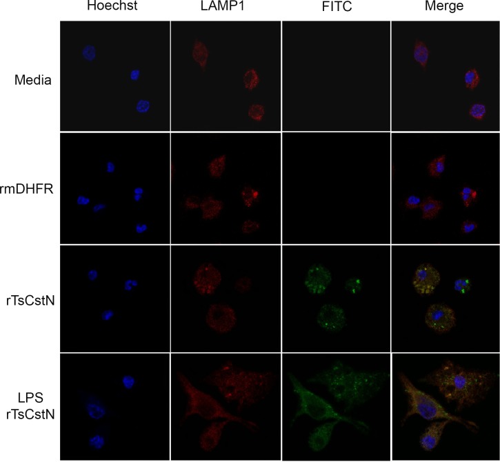
Fig 6. mBMDMs internalizing rTsCstN was determined using a laser scanning confocal microscope.
rTsCstN (green) were mainly detected in the cytoplasm of both untreated and LPS-treated mBMDMs. Moreover, rTsCstN were co-localized with CatL-containing lysosomes (yellow). Lysosomes were labeled with rabbit anti-human LAMP1 IgG plus Cy3-conjugated donkey anti-rabbit IgG (red), and the nucleus was counterstained with Hoechst 33342 (blue). Culture media- or FITC-labelled irrelevant control protein (rmDHFR) were used as negative controls.

