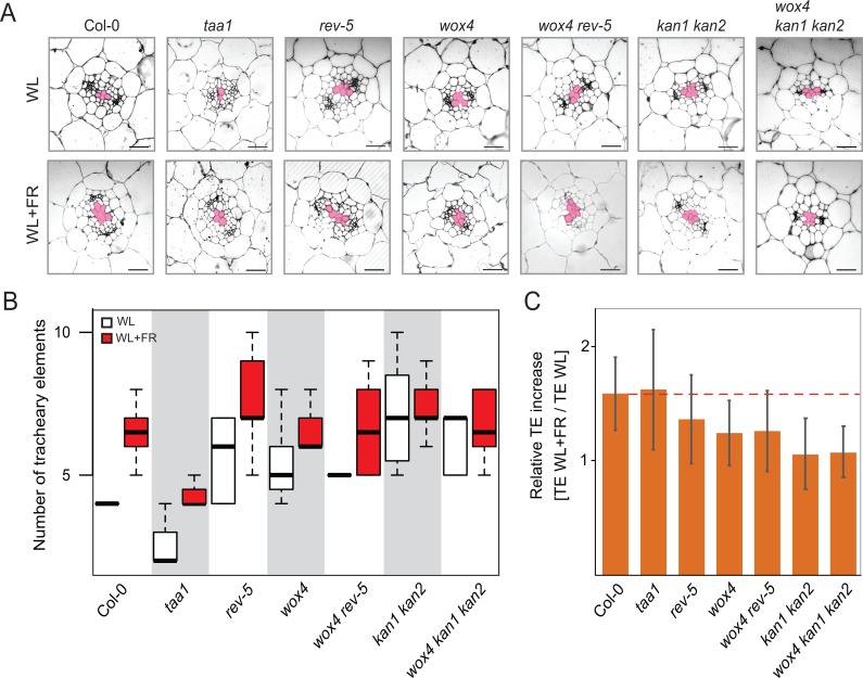
Fig 3. Shade-induced vascular patterning in mutants alters tracheary element formation.
A, Representative images of hypocotyl cross sections of 10-day old seedlings grown in white light (WL) or shade (WL+FR) conditions. Pink colored areas mark the TE cells in the center of the vascular cylinder. Scale bars, 20 μm. B, Box plots show the observed experimental data of TE numbers in WL and W+FR conditions for wild type and different mutants. Shown is the average of two biological replicates n = 9–13. Except taa1 which is based on one biological experiment n = 4–7. C, Relative TE increase by dividing the number of TEs in WL+FR with the number of TEs in WL. Red line indicates the ratio of the Col-0 wild type.

