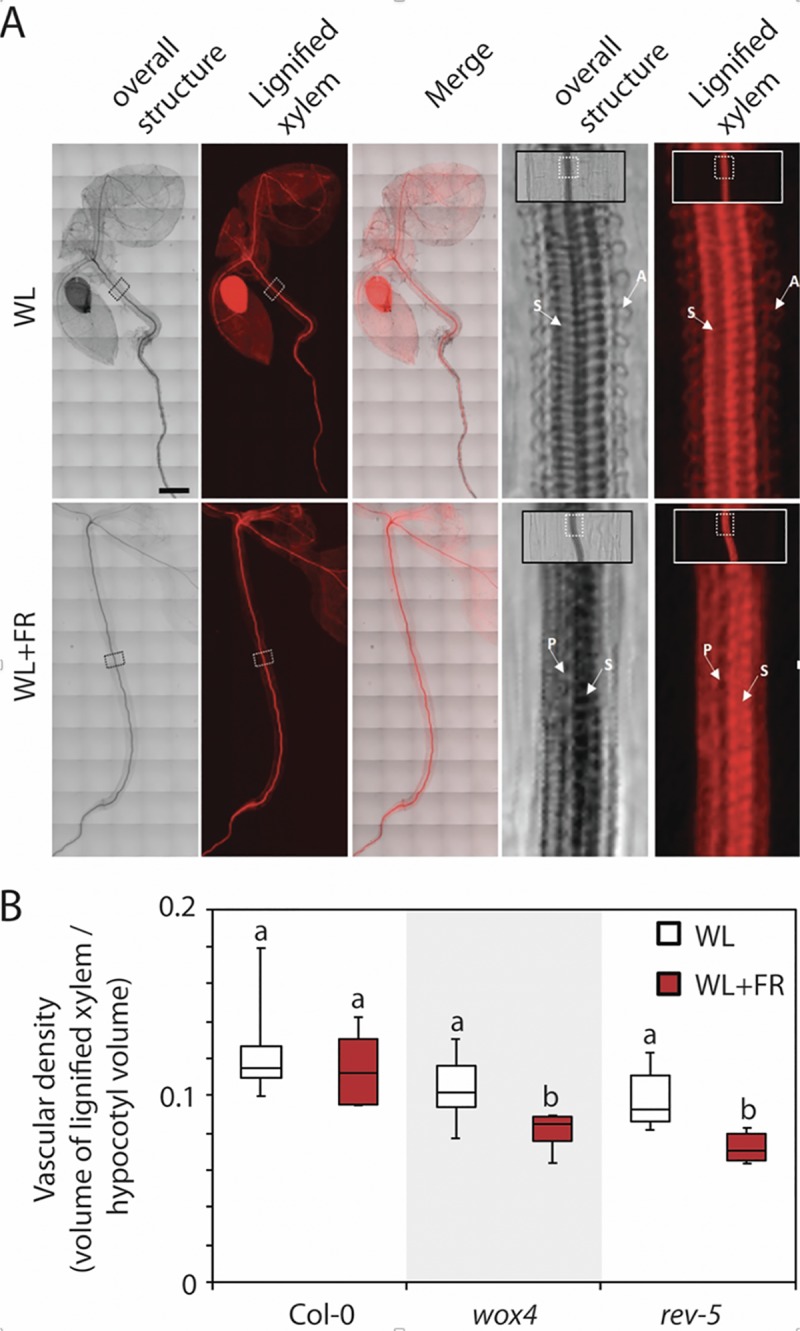
Fig 4. Virtual sections of white light and shade grown plants.

A, Whole seedling tomography experiment with Col-0 wild type plants. Square boxes in the whole seedling images are enlarged to the right to highlight the organization of the vascular system. Arrows indicate tracheary elements with different secondary cell wall patterning: A for annular, S for spiral and P for pitted. Scale bar = 250 μm. B, Measurements of the vascular density defined by the volume of the lignified xylem divided by the hypocotyl volume. (n = 6 seedlings per genotype).
