
Figure 5. Death by suicide does not appear to drive the differential PDE2A mRNA expression that was measured in patients with psychiatric illnesses.
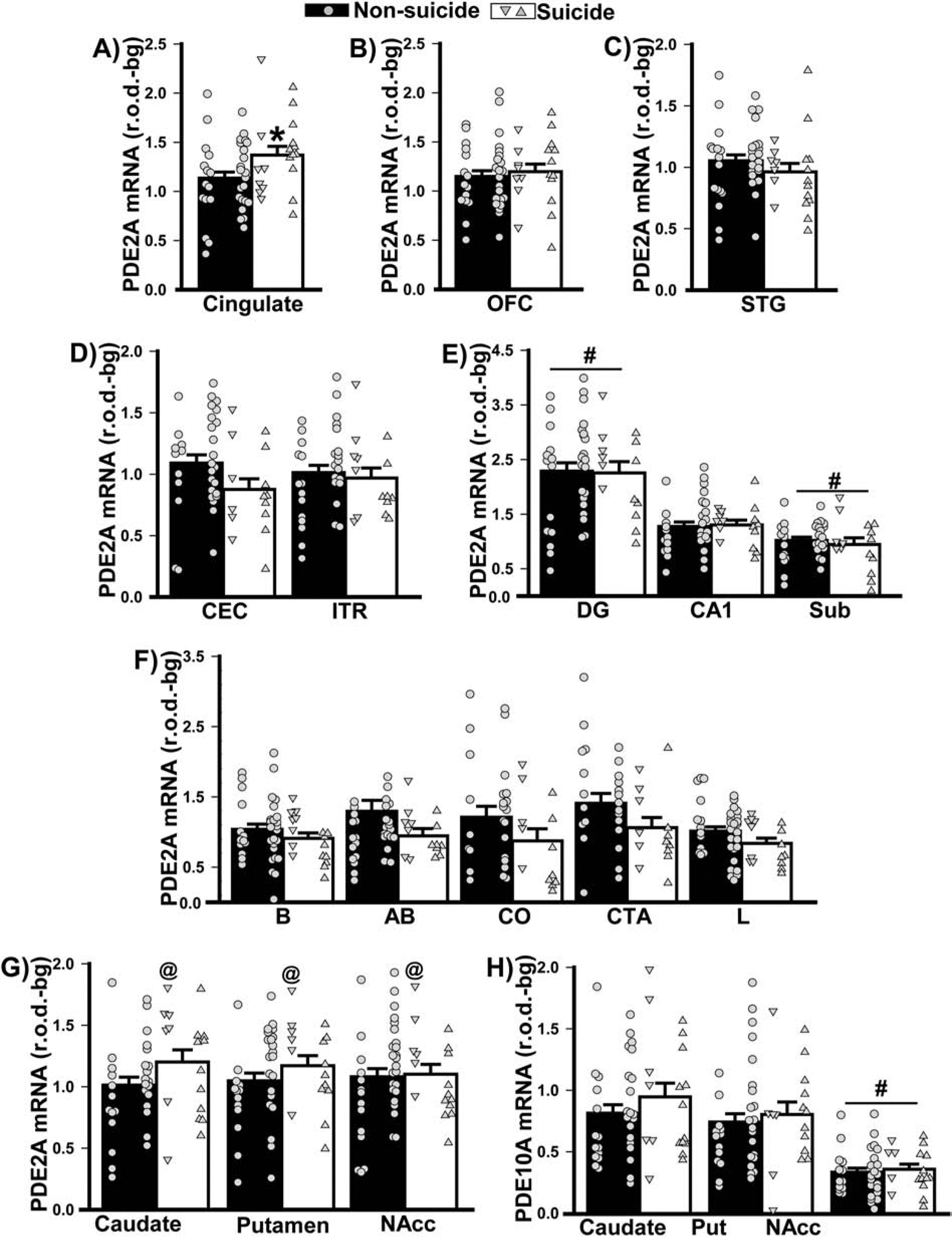
Data shown in Figure 2 were replotted based on mode of death (i.e., suicide versus non-suicide). A) In females (left data points) and males (right data points), death by suicide was associated with increased PDE2A mRNA expression in cingulate cortex (F(1,53)=4.33, P=0.042). No significant effects of mode of death were noted in B) orbitofrontal cortex (OFC), C) superior temporal gyrus (STG), D) parahippocampal cortices (i.e., caudal entorhinal cortex, CEC; inferotemporal region, ITR), E) hippocampus (including dentate gyrus, DG; subiculum, Sub), and F) amygdala (including basal nucleus, B; basal accessory nucleus, AB; corticalis nucleus, CO; cortico-amygdalar transition area, CTA; lateral nucleus, L). G) Across striatal nuclei (Putamen, Put; Nucleus Accumbens, NAcc), females who died by suicide expressed significantly higher PDE2A mRNA relative to females who died by other means (F(3,93)=5.04, P=0.0038; post hoc: Suicide female vs. non-suicide female, P=0.0067). H) In contrast, there was no significant effect of suicide in either males or females on striatal PDE10A mRNA expression. *vs. non-suicide, P=0.042; @vs. non-suicide female, P=0.007; # vs. other regions, P=0.0009–0.0001.