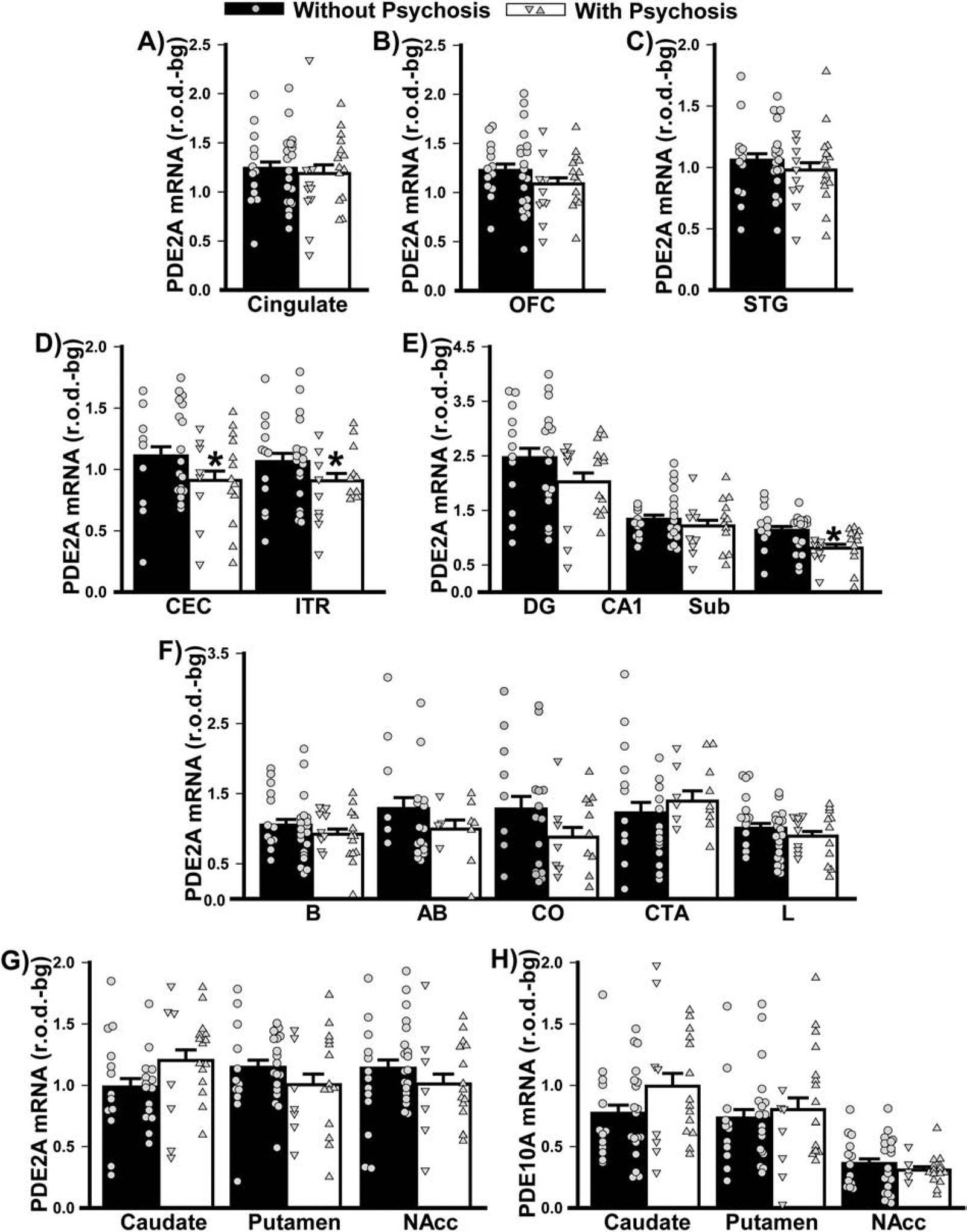
Figure 6. Psychosis cannot wholly account for the PDE2A mRNA expression that were noted in patients with psychiatric illnesses.

Data shown in Figure 2 were replotted based on history of psychosis. There was no significant difference between individuals with vs. without psychoses in terms of PDE2A mRNA expression in A) cingulate, B) orbitofrontal cortex (OFC), or C) superior temporal gyrus (STG). D) In contrast, individuals with vs. without psychosis expressed significantly lower PDE2A mRNA across the parahippocampal cortical subregions (F(1,44)=04.35, P=0.042) as well as E) subiculum (Sub; failed normality; T(20,28)=337.0, FDRP=0.0043). F) There were no significant effects of psychosis on PDE2A mRNA in the amygdala, G) PDE2A mRNA in the striatum (psychosis x region: F(2,97)=3.92, P=0.023; however, no post hoc tests reached significance), nor H) PDE10A mRNA in the striatum. *vs. without psychosis, P=0.042–0.004.
