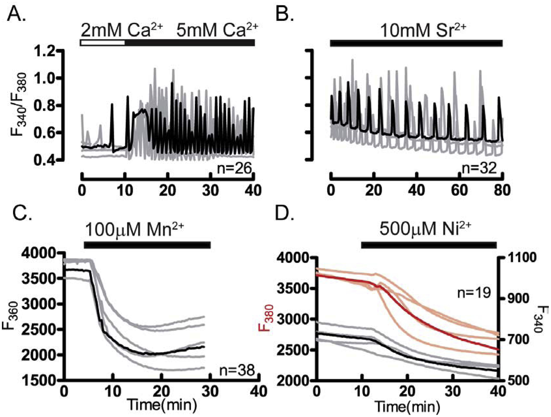
Figure 2. Divalent cations permeate the plasma membrane of GV oocytes.

Divalent cation influx was examined in GV oocytes. (A) Representative traces of Ca2+ oscillations caused by enhanced influx after increasing [Ca2+]o from 2 mM to 5 mM. (B) Sr2+ induced oscillations. (C) Fluorescence quenching at F360 following Mn2+ addition (100 μM). (D) Fluorescence quenching at F380 following Ni2+ addition (500 μM) in oocytes loaded with Mag-Fura-2AM in DFM media. Horizontal bars above each panel show the time during which divalent cations were added to the media. All experiments were replicated 4 times. In all panels in this figure and throughout the manuscript, representative traces are shown; darker traces represent the most common response; n: total number of oocytes examined.
