
Figure 3. Ca2+ influx in GV oocytes is mediated by multiple channels.
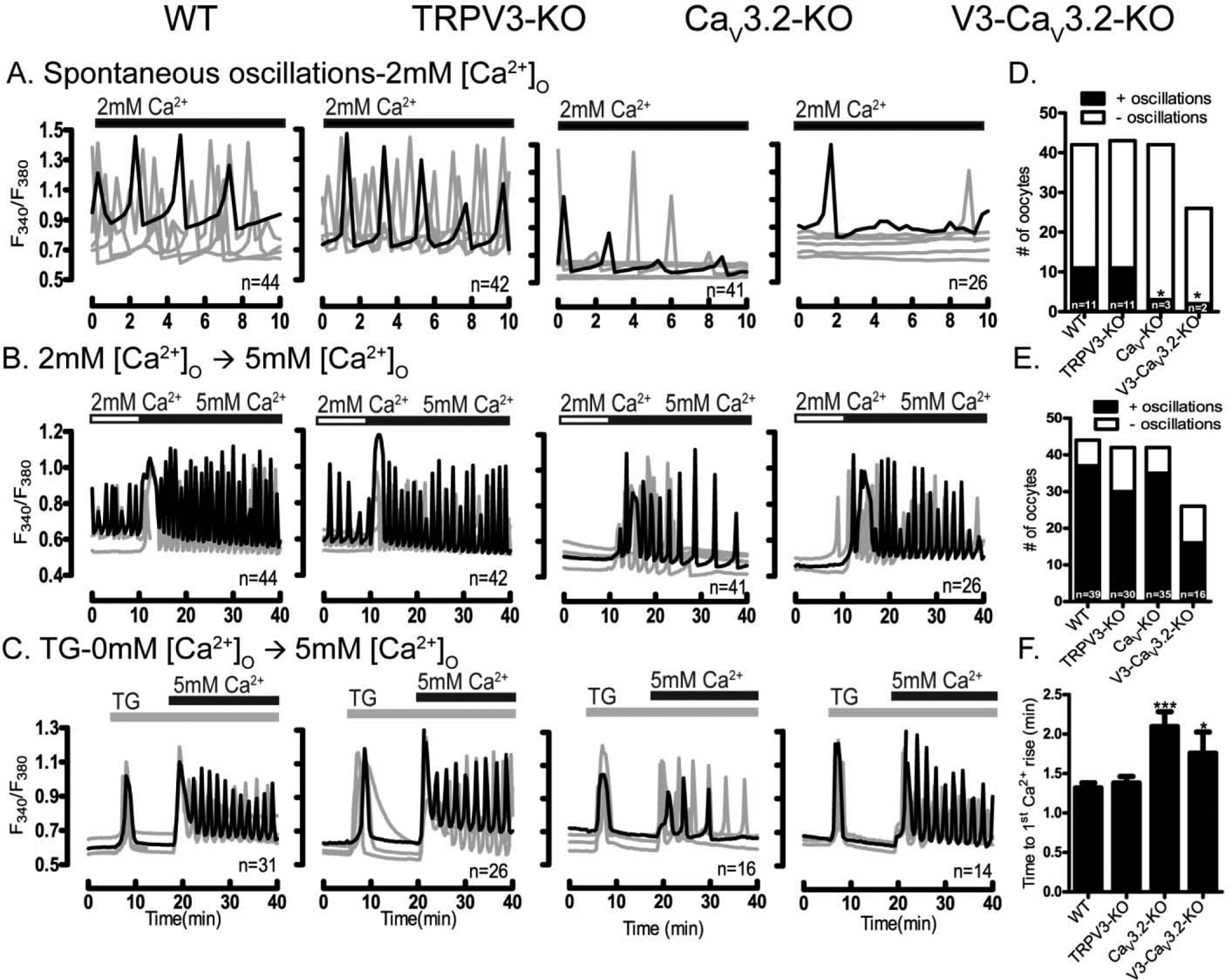
Presence of spontaneous oscillations and Ca2+ influx examined in WT oocytes and oocytes lacking TRPV3, CaV3.2 & V3-CaV3.2 channels. (A-four upper panels) Spontaneous Ca2+ oscillations in WT and KO oocytes in media containing basal Ca2+ levels (2 mM). Fewer oocytes of the CaV3.2 and V3-CaV3.2 KO lines display oscillations, which are also of decreased frequency (D) (P< 0.05). (B-four medium panels) Ca2+ influx triggered by increasing [Ca2+]o from 2 mM to 5 mM induces oscillations in all WT and KO lines, which appear of equal initial frequency (E). (C-four lower panels) After addition of TG (10 μM), Ca2+ influx resulted in reduced responses in CaV3.2 and V3-CaV3.2 KO oocytes, especially in the interval from Ca2+addition to the 1st Ca2+ peak (P< 0.05) (F). Filled horizontal bars above each panel show the time of addition of Ca2+ (black) or TG (grey) to the media. All experiments were 4 replicated for times. Asterisk(s) above columns in bar graphs denote significant differences from other conditions here and throughout the manuscript.