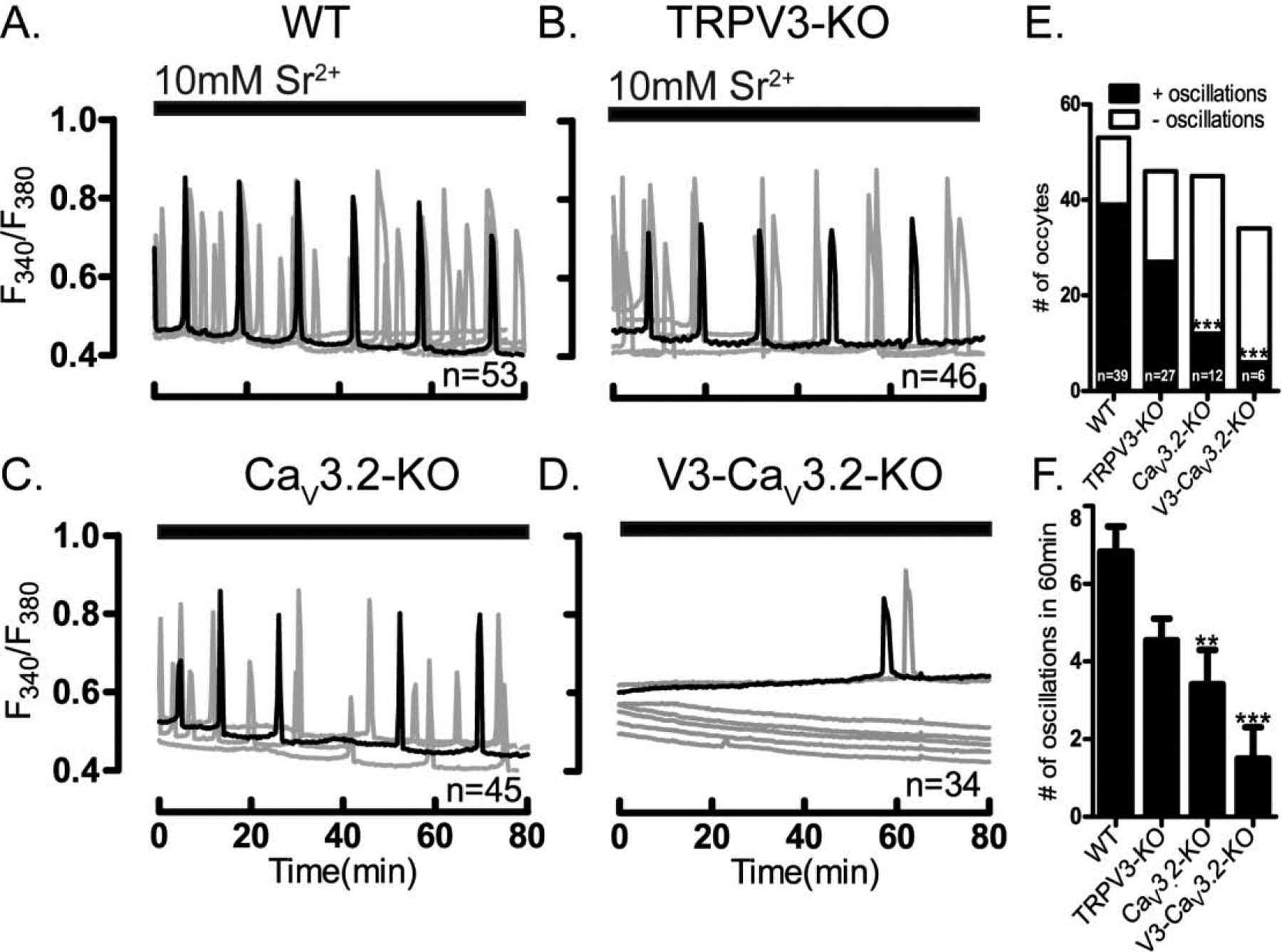
Figure 5. Sr2+-induced oscillations are diminished in CaV3.2- and V3-CaV3.2-KO oocytes.

Sr2+ induced oscillations were tested in WT and in oocytes lacking TRPV3, or CaV3.2 or V3-CaV3.2 channels. Experiments were performed in nominal Ca2+-free media containing 10 mM Sr2+. (A) Sr2+ induced the expected oscillations in WT oocytes, and largely similar responses in (B) TRPV3-KO oocytes. Sr2+ induced oscillations were reduced in (C) CaV3.2-KO and in (D) V3-CaV3.2-KO oocytes both in the (E) number of cells showing oscillations, and in the (F) number of rises during the first 1h of imaging (P<0.05). Horizontal bars above each panel denote the time during which Sr2+ was present in the media. All experiments were performed in 3–4 replicates.
