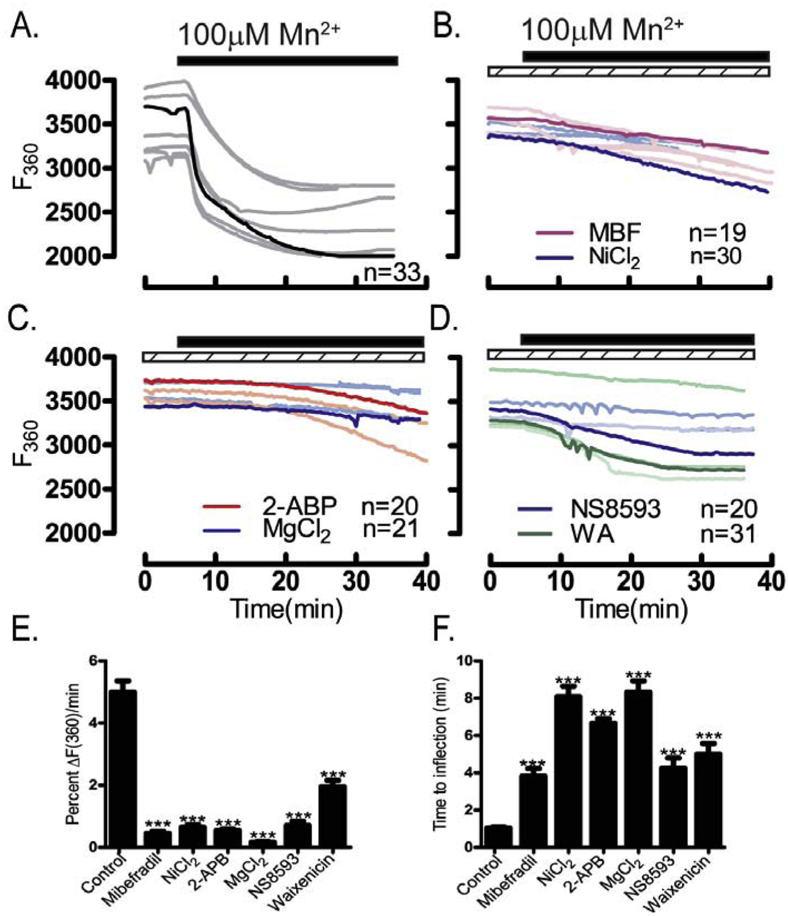
Figure 8. Inhibitors of CaV3.2 & TRPM7 channels suppress Mn2+ influx.

Mn2+ influx monitored using traces depicting quenching of F360 nm fluorescence. Experiments were performed in media containing 2 mM Ca2+ in the absence/presence of specific pharmacological inhibitors (dashed bars) after addition of Mn2+ (black bars): (A) control untreated oocytes, (B) oocytes treated with the CaV3.2 inhibitors MBF (1 μM) or Ni2+ (100 μM), (C) or with the general TRP inhibitors 2-APB (50 μM) and MgCl2 (10 mM), or (D) with the TRPM7 inhibitors NS8593 (10 μM) or waixenicin-A (1 μM). (E) Rate of fluorescence quenching per minute at F360 nm relative to baseline fluorescence was reduced by all inhibitors examined (P<0.05), and (F) time to inflection measured from the time of addition (5 min) to the time fluorescence quenching begins was prolonged by all inhibitors examined(P<0.05). Experiments were replicated 3 times.
