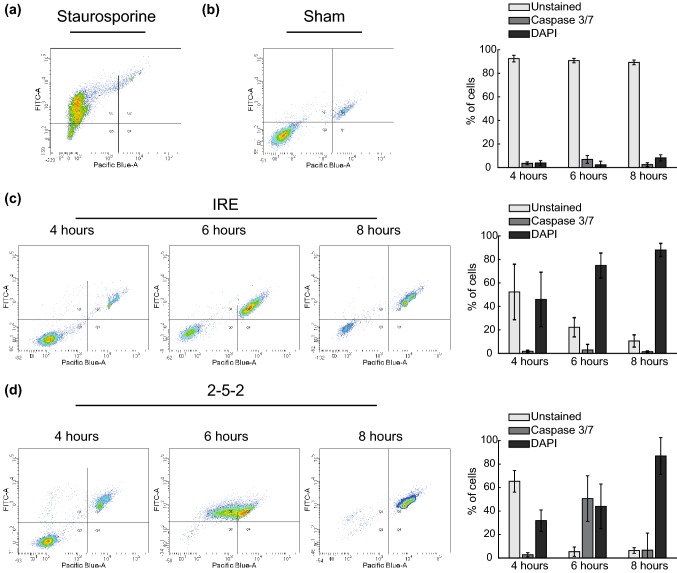
Figure 6.
Cells treated with H-FIRE exhibit increased Caspase activity and a sharp loss of membrane integrity vs. IRE-treated cells, which gradually internalize DAPI without Caspase activation. Assessment of Caspase 3/7 activity and membrane integrity after treating cells in suspension with IRE pulses of 1100 V/cm and 2-5-2 bursts with 3800 V/cm of amplitude. Representative examples of the density plots obtained with flow cytometry analysis are presented together with the time evolution of the percentage of cells in the different populations. DAPI fluorescence was measured in the Pacific Blue channel and CellEvent Fluorescence in the FITC channel. Cells were classified as: Unstained (bottom left corner), Caspase 3/7 positive (upper left corner) and DAPI positive (upper right corner). The values in the bar plots are presented as mean ± standard deviation (n ≥ 7 in all treatment groups and time points). (a) Density plot of cells treated with 1 μM Staurosporine for 8 h (positive control for Caspase 3/7 expression) (b) Density plot and time evolution of the Sham group. (c) Density plots and time evolution of IRE treated cells. (d) Density plots and time evolution of cells treated with 2-5-2 H-FIRE scheme.

