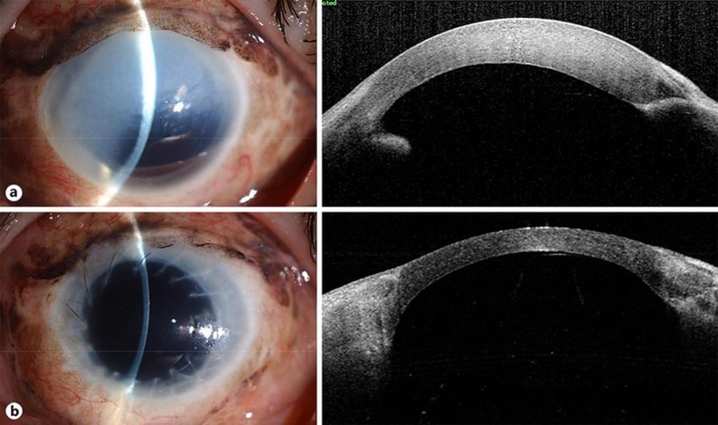
Fig. 2.
Anterior ocular segment images of the patient's left eye before (a) and 6 months after surgery (b). a Corneal clouding was apparent due to bullous keratopathy, and the cornea appeared thickened. Left and right images were obtained with a slitlamp microscope and by optical coherence tomography, respectively. b The transplanted cornea was clear and of normal thickness.

