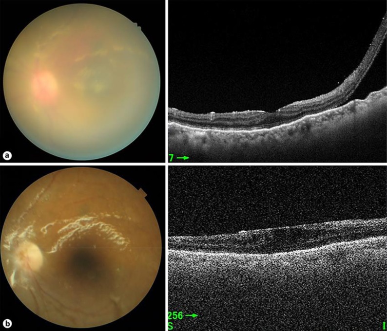
Fig. 3.
Posterior ocular segment images of the patient's left eye before (a) and 6 months after surgery (b). a Retinal detachment was apparent by optical coherence tomography (right), but it was difficult to perform funduscopy (left). b The transplanted cornea remained clear, so it was possible to perform funduscopy, and retinal redetachment had not occurred.

