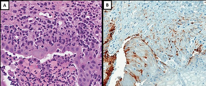
Fig. 3.
A Oral ulcer contained and surrounded by polymorphonuclear cells and confined cell debris; outside this zone and extending diffusely throughout the lamina propria, a large number of infiltrating lymphocytes and macrophage-like cells were seen (×40). B Inflammatory cell infiltrate (CD8+ stained; antibody CONFIRM anti-CD8, SP57) associated with oral mucosa ulceration (×10).

