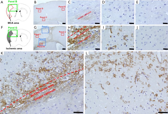
Figure 1.
CD44+ cells emerge within ischemic areas during acute phase following stroke. Immunohistochemistry at post-stroke day 7 (A-E) displays weak CD44 staining in the corpus callosum in sham-operated mice (B,C). CD44 was rarely observed in cells of MCA (B and D) and ACA regions (B and E). On day 7 after ischemic stroke (F-I), in addition to the increased expression of CD44+ cells in the corpus callosum (G and H), many CD44+ cells also emerged in peri-ischemic areas (G, H, and K) and ischemic cores (G, I, K, and L). In contrast, CD44+ cells were rarely observed in ipsilateral non-ischemic areas (G and J). Results displayed are representative of three replicates (N=3). Scale bars =200 µm (B and G), 20 µm (C, D, E, H, I, and J), and 50 µm (K and L). MCA, middle cerebral artery; ACA, anterior cerebral artery.

