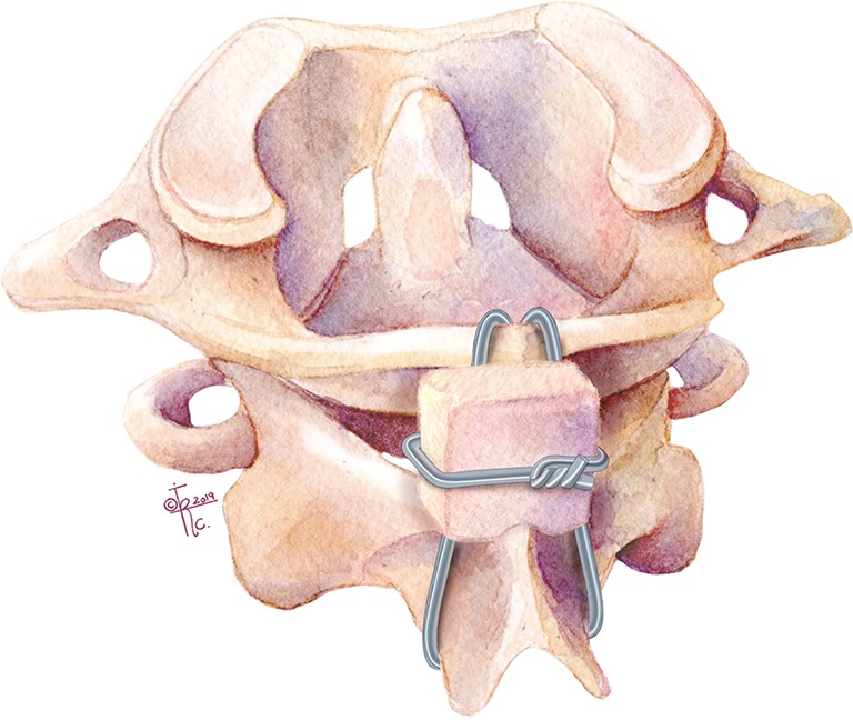
Figure 1.

Illustration of the Gallie sublaminar technique. Wire placed under C1 posterior arch, looping around the C2 spinous process, and then performing “on-lay” fusion by placing iliac crest bone with a midline notch to dock onto the C2 lamina and spinous process.
