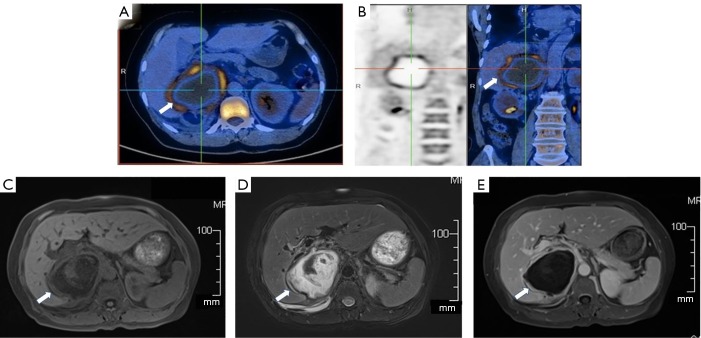
Figure 1.
PET/CT and abdomen magnetic resonance imaging. (A,B) Revealed a hypodense lesion in the right suprarenal region (arrows) with mild elevated SUVmax 6.7. (C,D,E) Showed right adrenal mass (arrows) with a thick wall and inhomogeneous signal inside. (C) T1-weighted image on admission; (D) T2-weighted image on admission; (E) T1-weighted image with contrast on admission.

