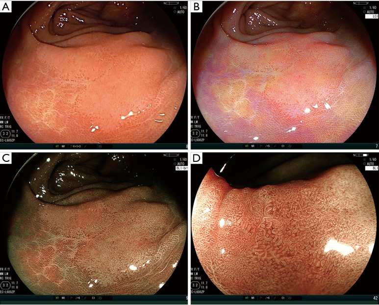
Figure 3.
A case presentation of LASER endoscope with linked color imaging (LCI) and blue laser imaging (BLI). (A) White light imaging. Non-polypoid lesion, 20 mm, Transvers colon. High-grade dysplasia; (B) LCI improves tumor detectability; (C) BLI improves tumor detectability; (D) BLI with magnification showed irregular pattern as JNET type 2B. JNET, Japan NBI Expert Team.

