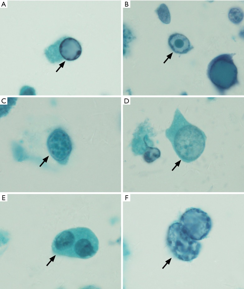
Figure 1.
On Papanicolaou staining (×1,000), the decoy cells exhibited 4 classical morphological types. (A) Type 1 classic decoy cell contained enlarged, basophilic, homogeneous amorphous ground glass-like intra-nuclear inclusion body (arrow); (B) type 2 decoy cell contained a single nuclear inclusion body surrounded by a peripheral halo, appearing as a bird’s eye showing (arrow); (C) type 3 decoy cell contained scattered clumped chromatin (arrow); (D) type 4 decoy cell contained cytoplasmic vesicles with fine-grained chromatin and nucleoli (arrow); (E) decoy cell contained two nucleus (arrow); (F) inclusion-bearing cell with degenerative changes in the cytoplasm, which appeared an apoptotic scenario (arrow).

