Figure 1.

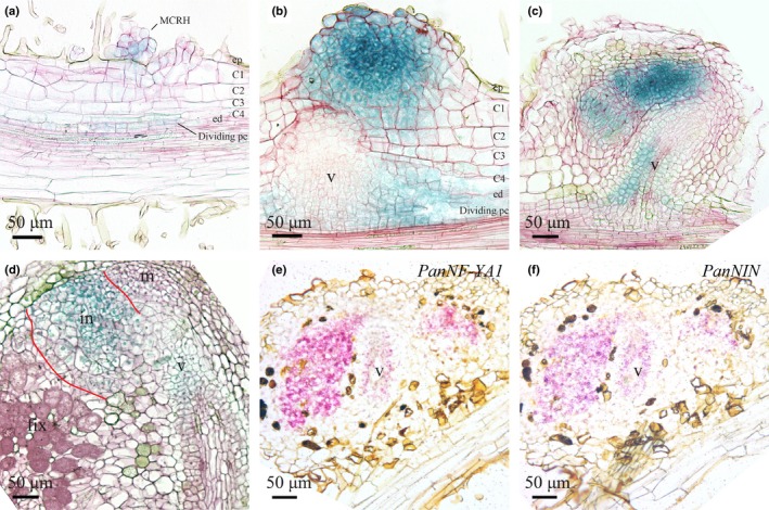
Spatiotemporal expression pattern of PanNF‐YA1 and PanNIN in developing Parasponia andersonii root nodules. (a–d) Spatiotemporal expression pattern of PanNF‐YA1pro:GUS in nodules of different developmental stages. (e, f) Spatiotemporal expression pattern of PanNF‐YA1 and PanNIN visualized by in situ hybridization on consecutive sections of a young P. andersonii nodule primordium. (a) PanNF‐YA1pro:GUS activity in clustered root hairs that are associated with dividing epidermal, outer cortical and pericycle cells. (b) PanNFYA1pro:GUS activity in a young but not yet intracellularly infected nodule and in the pericycle‐derived cells flanking the developing nodule vasculature. (c) PanNF‐YA1pro:GUS activity in the infection zone of young nodules, and in the basal part of the nodule vasculature. (d) PanNF‐YA1pro:GUS activity in a mature nodule is restricted to the infection zone (marked with red lines) and nodule vasculature. (e, f) PanNF‐YA1 (e) and PanNIN (f) transcripts are detected in the infection zone and nodule vasculature by in situ hybridization on consecutive sections. MCRH, multicellular root hairs; ep, epidermis; C1‐C4, first to fourth cortical cell layer; ed, endodermis; pc, pericycle; m, nodule meristem; in, infection zone; fix, fixation zone; v, nodule vasculature. In (a)–(d), sections (7 µm) were counterstained with Ruthenium Red. Nodules were isolated 4 wk post‐inoculation with Mesorhizobium plurifarium BOR2.
