Figure 5.

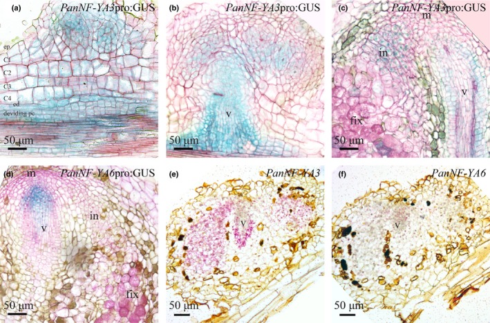
Spatiotemporal expression pattern of PanNF‐YA3 and PanNF‐YA6 in Parasponia andersonii root nodules. (a, c) Spatiotemporal expression pattern of PanNF‐YA3pro:GUS in nodules of different developmental stages. (a) PanNF‐YA3pro:GUS activity is observed in dividing epidermal, cortical, endodermal cells of a nodule primordium as well as the root vasculature. (b) In a young nodule, PanNF‐YA3pro:GUS activity is confined to the nodule lobes that will become intracellularly infected and the nodule vasculature. (c) In a mature nodule PanNF‐YA3pro:GUS is active in the infection zone and the nodule vasculature (v). (d) PanNFYA6pro:GUS is active at the nodule vascular meristem. (e, f) Spatiotemporal expression pattern of PanNFYA3 and PanNF‐YA6 visualized by in situ hybridization on consecutive sections of a young P. andersonii nodule primordium. ep, epidermis; C1–C4, first to fourth cortical cell layer; ed, endodermis; pc, pericycle; m, nodule meristem; in, infection zone; fix, fixation zone; v, nodule vasculature. In (a)–(d), sections (7 µm) were counterstained with Ruthenium Red. Nodules were isolated at 4 wk post‐inoculation with Mesorhizobium plurifarium BOR2.
