
Figure 4.
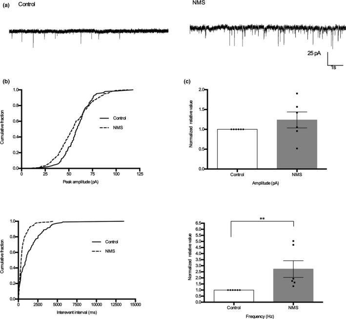
Synaptic activity of SDH neurons increased in NMS rats. (a) Representative traces of sEPSCs recordings from thoracolumbar spinal cord slices taken from control and NMS rats. (b) Cumulative fraction of peak amplitude and inter‐event interval of control and NMS rats. (c) Bar graph of amplitude and frequency normalized to the control rats. Neurons, n = 6, animals, n = 5, per group; **p < .01, t‐test