
Figure 1.
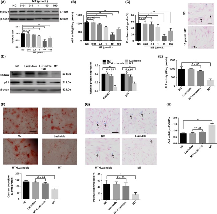
Melatonin can antagonise VSMC osteogenic differentiation and senescence via an MT membrane receptor‐dependent manner. A‐C, Effects of melatonin (MT) on RUNX2 expression, ALP activity and senescence level. VSMCs were treated with 10 mM β‐glycerophosphate (β‐GP) and different concentrations of MT (10 nM, 100 nM, 1 μM, 10 μM and 100 μM) or vehicle for the control. A, The expression of RUNX2 was determined by Western blotting. B, ALP activity was measured using an ALP kit, normalised to the cellular protein content. C, SA‐β‐gal staining was performed to detect cell senescence level. Semi‐quantitative analysis of SA‐β‐gal–positive cells (left panel) and representative microphotographs (right panel) are shown. D, The protein levels of RUNX2 and p21 were determined by Western blotting in vehicle, luzindole, 10 μM MT plus luzindole or 10 μM MT‐treated VSMCs. E, ALP activity assays were measured using an ALP kit for vehicle, luzindole, 10 μM MT plus luzindole or 10 μM MT‐treated VSMCs. F, β‐GP–induced VSMCs were treated with vehicle, luzindole, 10 μM MT plus luzindole or 10 μM MT for 14 days and then subjected to Alizarin Red S staining. Calcium deposition was extracted with cetylpyridinium chloride and quantified by spectrophotometry. G, VSMCs were treated with vehicle, luzindole, 10 μM MT or 10 μM plus luzindole and then subjected to SA‐β‐gal staining. Semi‐quantitative analysis of SA‐β‐gal–positive cells was performed. Representative microscopic views are shown. Scale bar represents 200 µm. H, CCK8 assay was performed in VSMCs treated with vehicle, luzindole, 10 μM MT plus luzindole or 10 μM MT. The data represent the mean ± SD. NC, normal control. *P < .05, **P < .01