
Figure 4.
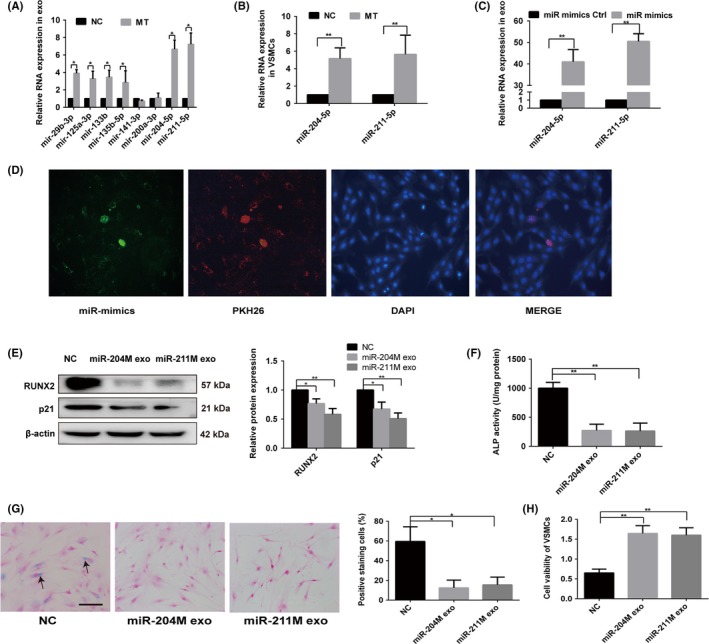
MT induced exosomal miR‐204 and miR‐211 antagonise VSMC osteogenic differentiation and senescence. A, qRT‐PCR analysis of eight microRNAs in exosomes derived from MT‐treated VSMCs or vehicle‐treated VSMCs. B, qRT‐PCR analysis of miR‐204 and miR‐211 expression in VSMCs treated with MT or vehicle. C, qRT‐PCR analysis of miR‐204 and miR‐211 in VSMCs after miR‐204/211 mimics or mimics control transfection. D, Confocal microscopy analysis was used to verify exosomal miR‐204 could be uptaken by VSMCs. VSMCs were cultured with PKH26‐labelled exosomes derived from VSMCs transfected FAM‐miR‐204‐mimics. The FAM‐miR‐204 signals were detected in the cytoplasm of VSMCs (Green), and FAM‐miR‐204 signals were co‐localised with PKH26 in VSMCs (PKH26 in red, DAPI in blue). Magnification, 100×. E, The expression of RUNX2 and p21 was determined with Western blotting in VSMCs treated with exosomes from miR‐204/211 overexpression VSMCs or exosomes from mimic control transfected VSMCs. F, ALP activity was determined using an ALP kit in VSMCs treated with exosomes from miR‐204/211 overexpression VSMCs or exosomes from mimic control transfected VSMCs. G, VSMCs treated with exosomes from miR‐204/211 overexpression VSMCs or exosomes from mimic control transfected VSMCs and then subjected to SA‐β‐gal staining. Semi‐quantitative analyses of SA‐β‐gal–positive cells were performed. Representative microscopic views are shown. Scale bar represents 200 µm. H, CCK8 assays were performed in VSMCs treated with exosomes from miR‐204/211 overexpression VSMCs or exosomes from mimic control transfected VSMCs. Three independent experiments were performed, and representative data are shown. NC, normal control. *P < .05. **P < .01. NC, normal control