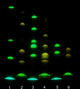
Figure 2.

Gel electrophoresis (migration from “north” to “south”, pH 8.3); detection by emission (excitation at 365 nm). From bottomn to top. Lane 1: APTS (lowest; blue), APTS+G, APTS+G3, APTS+G7 (green). Lane 2: dye 16 (green), 16+G, 16+G3, 16+G7 (yellow). Lane 3: APTS, APTS+M, APTS+M2‐2O/APTS+M2‐3O (unresolved), APTS+M2‐4O, APTS+M3 and APTS+M4. Lane 4: dye 16, 16+M, 16+M2‐3O, 16+M2‐2O/16+M2‐4O (unresolved), 16+M3 and 16+M4. Lane 5: APTS, APTS‐labeled 3′‐ and 6′‐sialyllactoses. Lane 6: dye 16 and its conjugates with 3′‐ and 6′‐sialyllactoses.
