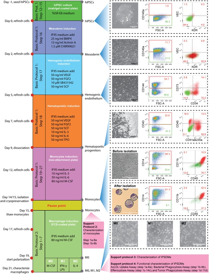
Figure 1.

Timeline of all protocol procedures. Bright‐field images of representative cellular morphology are shown for day 0 (undifferentiated hiPSCs), day 2 (mesoderm), day 5 (HE), day 9 (HPCs), day 14 or 15 (monocytes before isolation), and day 21 (M0, M1, and M2 macrophages). FACS analysis of stage‐specific markers at day 0, day 2, day 5, day 9, and day 15 of differentiation are shown. Wright‐Giemsa staining of hiPSC‐mono after isolation at day 15 is also shown. Scale bar represents 200 μm in all bright‐filed images and 50 μm in Wright‐Giemsa staining. Abbreviations: hiPSCs, human induced pluripotent stem cells; HE, hemogenic endothelium; HPCs, hematopoietic progenitor cells; FACS, fluorescence activated cell sorting; hiPSC‐mono, hiPSC‐derived monocytes
