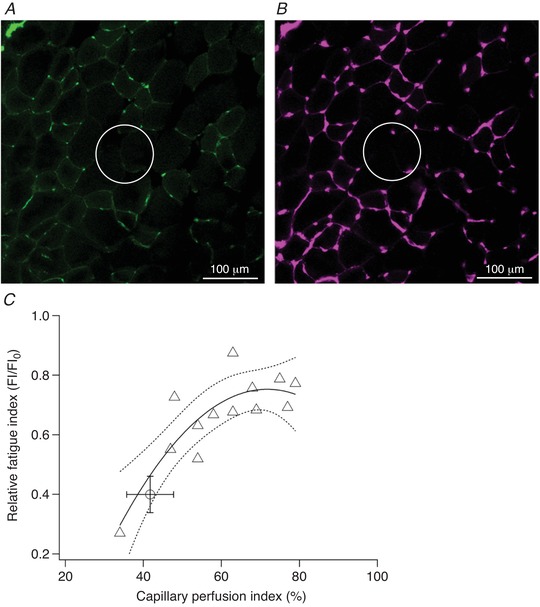
Figure 4. Histological quantification of microsphere‐injected extensor digitorum longus (EDL).

Dextran‐FITC‐labelled perfused (A) and total (B) capillary supply after moderate microsphere injection. Comparison of circled regions highlights functional microvascular rarefaction as capillaries remain unperfused after arteriolar blockade. There was a significant polynomial relationship (r 2 = 0.720, P = 0.002) between ipsilateral capillary perfusion index (i.e. proportion of total capillaries that were perfused) and relative fatigue index (FI) in animals undergoing acute injection of microspheres (C). The effect of femoral ligation is also shown for comparison (open circle). Regression line and 95% confidence intervals are plotted.
