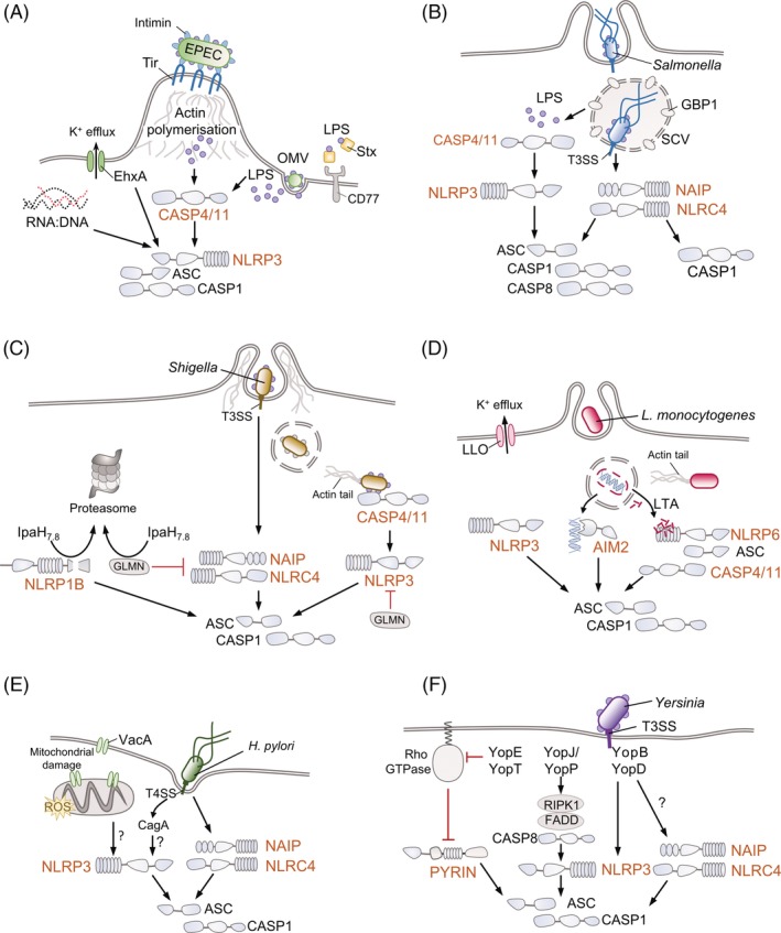
Figure 2.

Enteric pathogens can activate multiple inflammasome pathways. Schematics from (a) to (f) show how various enteric pathogens stimulate the assembly and activation of different inflammasomes, focussing on the activating signal (signal 2). Downstream consequences of inflammasome activation shown in Figure 1, that is Gasdermin D cleavage, pore formation and pyroptosis, caspase‐1‐mediated cleavage of pro‐IL‐1β and pro‐IL‐18 into their active forms and the release of these pro‐inflammatory cytokines together with alarmins, are not depicted for simplicity. A/E pathogens (such as Enteropathogenic and Enterohaemorrhagic E. coli, EPEC and EHEC) (a), Helicobacter pylori (e) and Yersinia (f) are mainly extracellular pathogens, while Salmonella survives intracellularly in Salmonella containing vacuoles (SCVs) (b) and Shigella (c) and Listeria monocytogenes (d) are cytosolic bacteria that escape the vacuoles and can move from cell to cell via manipulation of the host actin cytoskeleton. Host proteins are featured in greyscale to emphasise the role of bacterial factors. See text for further information on the mechanisms employed by these pathogens to evade or subvert inflammasomes. OMV, outer membrane vesicle; Stx, Shiga toxin; Tir, translocated intimin receptor; GBP1, guanylate binding protein 1; GLMN, glomulin; LLO, listeriolysin; LTA, lipoteichoic acid; VacA, vacuolating cytotoxin A; CagA, cytotoxin‐associated gene A; ROS, reactive oxygen species; KD, kinase domain; RHIM, RIP (receptor‐interacting serine/threonine‐protein) homotypic interaction motif; DD, death domain; Yop, Yersinia outer protein
